
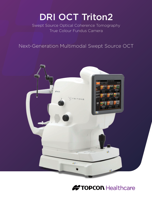
The New Generation Multimodal Swept Source Imaging Technology with DRI TRITON -2 Optical Coherence Tomography (OCT)
- Swept-Source OCT providing high density scans and deep penetration
- Slit-scan technology to image through small pupils (φ2.0mm or larger*1)
- Wide-field OCT and OCTA, up to 21mm*2
- Smart Denoise*2 provides higher signal-to noise ratio on 3D OCT and OCTA*2
- Flexible positioning for easier acquisition
- Simplified workflow with seamless integration for quick analysis and follow-up
Invisible Scan Lines
The invisible 1,050nm wavelength light helps patients concentrate on the fixation target during the scan, reducing involuntary eye movement. It supports more efficient workflow in practice by reducing the need to rescan.
Swept Source OCT Technology
A fast-scanning speed of 100,000 A-scans/sec enables capture of a dense array of clear B-scans by acquiring more A-scans within a given image acquisition time. This helps to reduce artifacts from involuntary eye movements such as saccades and blinks.
Combination Scan
The Triton2 allows for the combination of a 3D volumetric scan with reference database, and a high-resolution linear scan in a single acquisition.
Topcon’s optional SS OCT Angiography
The optional Topcon’s SS OCT Angio™ integrates OCT Angiography with Swept Source technology, and the long 1050 nm wavelength. Powered by OCTARA™, a proprietary image processing algorithm, SS OCT Angiography enables detailed visualization of vascular structures and the monitoring of key retinal pathologies.
Wide-field Imaging
The optional wide-field attachment lens enables the capture of scans up to 21mm in length. Gather more clinical insights with wide-field OCT and OCTA imaging – valuable in a wide variety of conditions.
OCTA Metrics
SS OCT Angio™ displays OCTA density which is the ratio between high and low signal areas. The information is displayed as a colour map with the ability to display values for rapid comprehension.
En Face OCT Imaging
En Face imaging allows for independent dissection and examination of key layers, such as the vitreoretinal interface (ILM boundary), retinal pigment epithelium and choroidal layers
Introducing New Slit-Scan Photography
The innovative slit-scan illumination and rolling shutter mechanism in the Triton2 produces excellent quality color fundus images with less flare and shadow. *3 The slit-scan mechanism helps to overcome one of the known causes of poorly graded images, with its ability to effectively image through small pupils.
This innovative technology also helps the Triton2 capture sharp, high-quality fundus images, regardless of miosis and the lighting conditions, unlike conventional fundus cameras.